Eyes
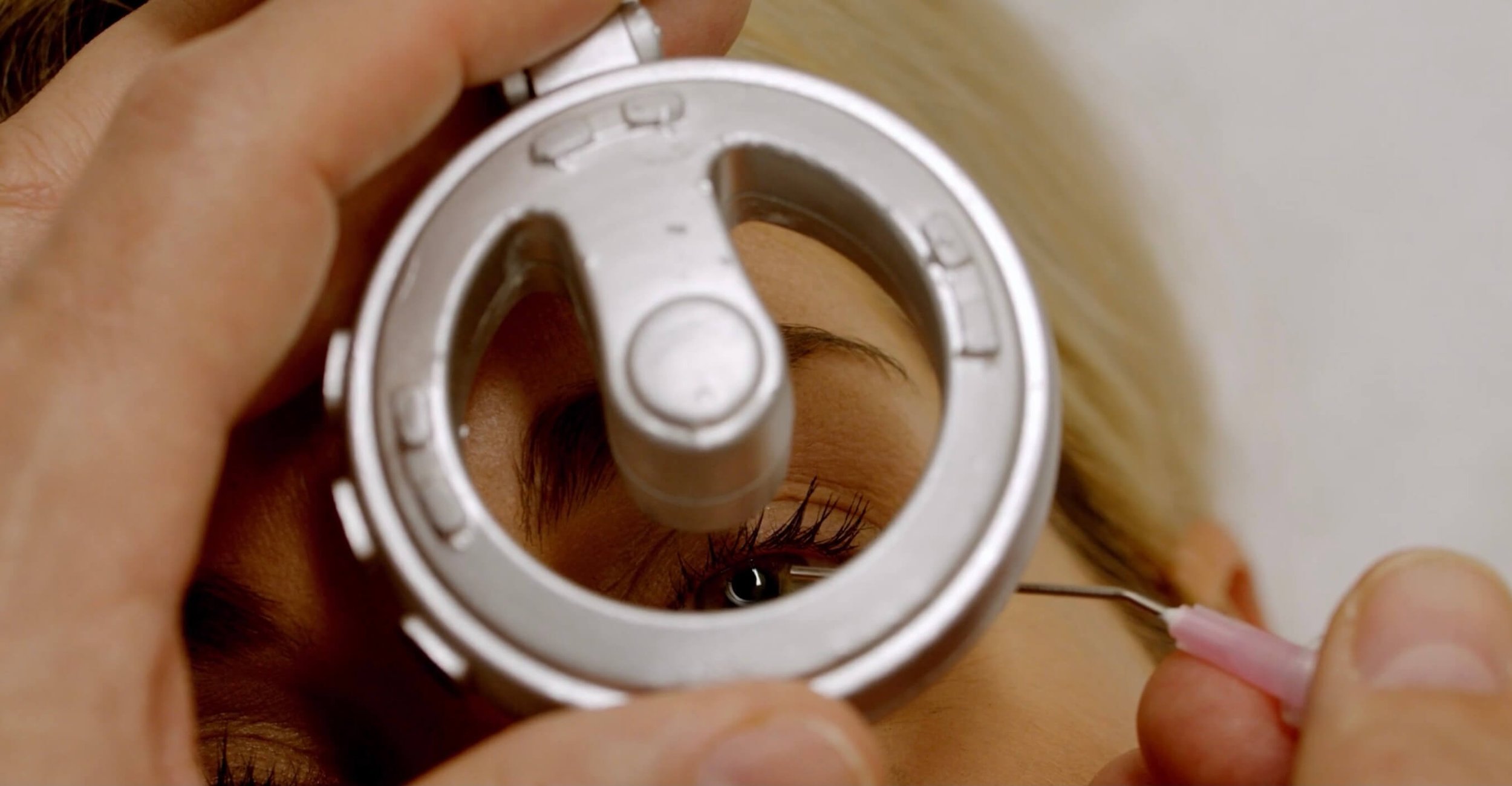
Inspection of the anterior eye, lens and the retina can be done at an arm’s length distance. It also has blue light and can be used together with fluorescein to assess corneal changes. Additionally, the iXope provides magnified visualisation for foreign body removal from the cornea.
Ophthalmoscope and Slit Lamp
iXOPE
iXope is a digital ophthalmoscope which supports general practitioners in performing billable eye procedures. Doctors are able to see eye, patient and operating hand at the same time which helps them having better coordination of their hand movement during procedures and noticing discomfort of the patient. Additionally, iXope’s software helps documenting and monitoring patients’ condition for acute or chronic disease management.
-
Unlike current slit lamps, where the doctor must examine through binoculars, iXope provides a magnified view of both the patients eye and the operating hand at the same time. With its large magnification, iXope can be used for detailed examinations of different structures of the eye like the cornea, lens or anterior chamber. iXope’s software also helps to document and monitor the patient’s condition as part of chronic disease management.
-
Using iXope, doctors are able to inspect the back of the eye (retina) for chronic disease management and acute eye conditions. Inspecting the retina with ophthalmoscopes requires specific skills and doctors have limited time to make their assessment during the examination of the retina. For these reasons, general practitioners often refer to optometrists or ophthalmologists, which is of course, more time-consuming and costly for the patient. The iXope combats this issue, allowing similar views of the retina, which is achieved by high-end ophthalmoscopes.
Doctors with glasses might not be able to use a conventional ophthalmoscope. iXope is used at a arm’s length distance making it comfortable for doctor and patient. Doctors with glasses will find it easy to use iXope as they look on to the device instead of through it.
Additionally, images can be reviewed without rush and be stored on the EMR or directly send to a specialists.
-
Each year over 2 million people across U.S., Europe and Australia end up at emergency departments presenting issues with foreign bodies in the eye. This costs the public health system around AU$1 billion!
Removing foreign bodies from the eyes requires a high-level of magnification and previously, a slit lamp which is only available with a specialist or in the hospital setting. However, majority of general practitioners have adequate training in this space. Therefore, iXope enables general practitioners to undertake these procedures in their own practice, giving patients access to instant treatment. Moreover, shifting this procedure from hospitals to general practitioners could save 85% of service hours and expenses for the healthcare system.
-
iXope can be effectively employed to help manage and treat chronic diseases related to the eyes such as macular degeneration and diabetic retinopathy. In acute cases, it can also be used to help doctors to detect retinal detachment, corneal infections or injuries, increased cranial pressure, haemorrhage or an acute occlusion of a retinal artery.
iXope has green light to filter the red colour which can assist in examining the retina. Thanks to the quality images provided via iXope, doctors can take their time establishing the diagnosis and attach the images to referrals. This allows patients to be managed and treated by their family doctor and will improve patient outcomes due to the high-quality documentation and improved collaboration provided by iXope.


